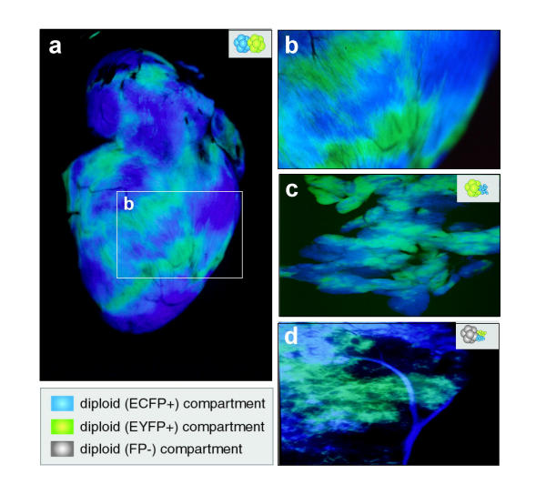
Figure 4.
Dual non-invasive reporter visualization in the organs of adult chimeras. All images depict dark field double exposures acquired by consecutively using ECFP and EYFP filters under dark field epifluorescence. Details of aggregates are schematized at the top right of each panel. (a-c) heart and pancreas from double-tagged double-compartment adult chimeras. (a) view of the surface of an adult heart. (b) close-up surface view of ventricle of a chimeric heart generated by aggregation of two diploid morulae, one hemizygous for the CK6/ECFP (ECFP+) transgene and the other hemizygous for the YC5/EYFP (EYFP+) transgene. The regions of yellow/green and cyan fluorescence are clearly mutually exclusive. The striations in the fields of fluorescence represent the proliferative zones present within the ventricle. (c) chimeric pancreas generated by aggregation of a morula hemizygous for the YC5/EYFP transgene with CK6/ECFP ES cells. The restricted fields of fluorescence represent the proliferative zones present within the pancreas. (d) liver from a double-tagged triple-compartment adult chimera. A non-fluorescent morula was aggregated with two clumps of ES cells, one carrying the CK6/ECFP transgene (ECFP+) and the other carrying the YC5/EYFP transgene (EYFP+). An ECFP+ vessel has clonally proliferated and infiltrated the liver lobe (top). Note that unlike in heart and pancreas there appears to be more interspersed blue/cyan vs green/yellow fluorescence present in the liver, possibly reflecting greater cell intermingling during the genesis of this organ.

