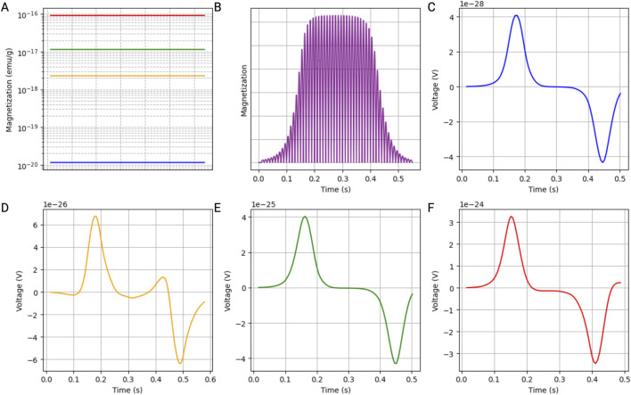
FIGURE 3.
Simulation results of a single iron oxide magnetic nanoparticle (MNP) within a blood vessel. The parameters were selected to accurately represent the rheological properties of blood under physiological conditions. (A) Magnetization of the MNPs, illustrating how different MNP sizes (5 nm: blue; 30 nm: yellow; 50 nm: green; 100 nm: red) respond to an alternating magnetic field. Those results were extrapolated from the M-H curves. (B) Magnetization profile of the MNPs over time, highlighting the oscillatory behavior induced by the alternating magnetic field, with the magnetization curve indicating the time at which the MNPs travel through the center of the coil. (C–F) Detailed voltage measurements over time, captured by the search coil system at different points within the simulated blood vessel, showing the induced signal by a single MNP with a diameter of 5 nm (C), 30 nm (D), 50 nm (E), and 100 nm (F).

