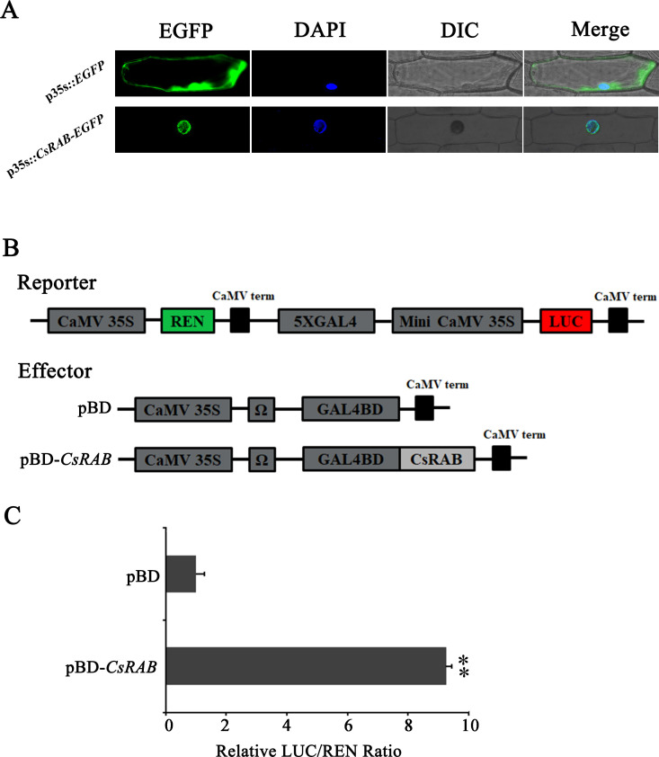
Figure 5.
Subcellular localization and transcriptional activation capability of CsRAB. (A) Subcellular localization of the CsRAB protein in onion (Allium cepa) epidermal cells. For each panel, EGFP: the enhanced green fluorescent image, DAPI: the 6-Diamidino-2’-phenylindole stained image, DIC: the bright field image, Merge: overlaid green fluorescence, DAPI stained and bright-field three images. The images show the nuclear localization of CsRAB. (B) The dual renilla luciferase (REN)/luciferase (LUC) reporter and effector constructs. The double-reporter plasmid contained 5× GAL4 and mini-CaMV35S fused to LUC and REN driven by CaMV35S. The effector plasmid contained the CsRAB gene fused to GAL4BD driven by CaMV35S. (C) The transcription activation ability of CsRAB. The LUC and REN luciferase activities were assayed after the dual REN/LUC reporter and effectors co-transformed into Arabidopsis protoplasts 24 h, and the transcription activation capability of CsRAB is indicated by the LUC to REN ratio. Each value represents the means of three biological replicates, and the asterisk indicates a statistically significant difference as assessed by Student’s t-test (∗∗P < 0.01) compared with pBD.

