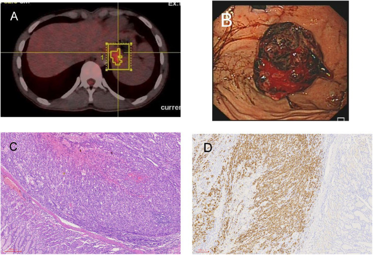
Figure 3.
Radiological and pathological images of left adrenal gland and stomach lesions. (A) Newly detected tumor in the left adrenal gland shown in PET-CT. (B) The tumor grew from the fundus into the gastric cavity, and the gastric stromal tumor ruptured and bled. (C) Stomach H&E staining. (D) Stomach immunohistochemistry: hepatocyte (+).

