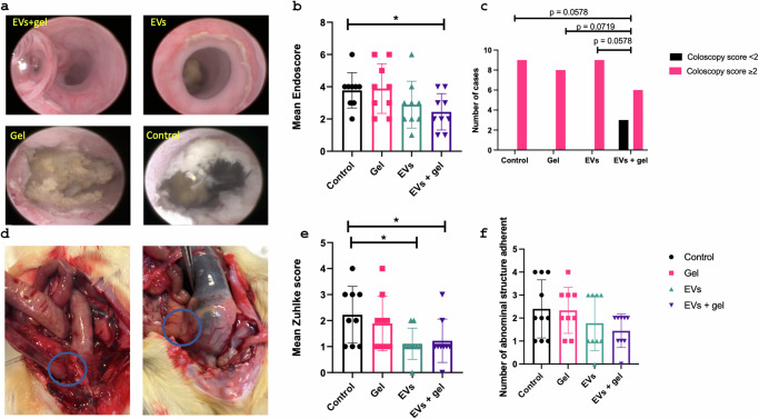
Fig. 5. Clinical evaluation at W8.
Representative colonoscopy pictures of the different group (a): the presence of anastomotic ulceration and its characterization was noted according to its severity (1: no ulcer, 2: superficial ulcer, 3: deep ulcer, 4: deep circumferential ulcer). Bleeding or stenosis of the anastomosis was also reported and combined in an ENDOSCORE (b and c). Pictures of colo-colic anastomosis during the autopsy (d). The blue circles highlight two entero-anastomotic fistulas. Graph representing the peritoneal adhesions by the Zuhlke score (e) and the number of structures adherent to the anastomosis (seminal vesicles, bladder, small bowel) (f).

