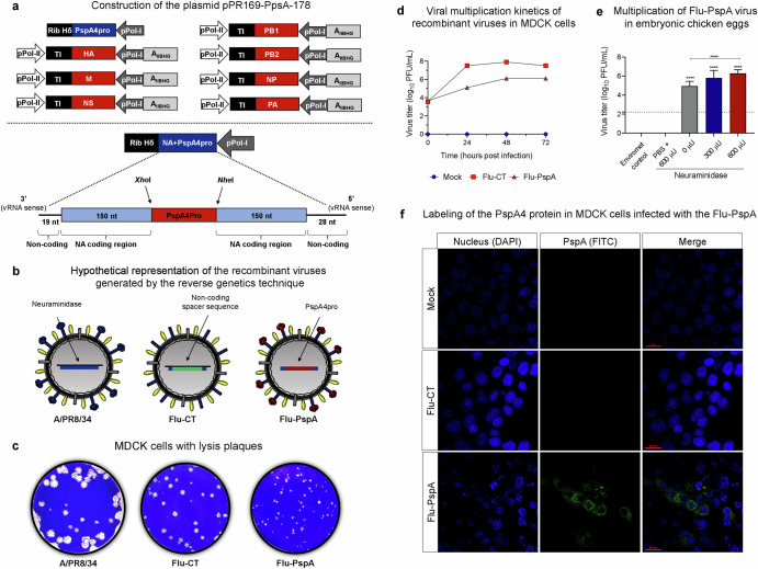
Fig. 1. Construction and characterization of recombinant Influenza virus Flu-PspA.
a Schematic representation of the plasmids that encode the segments of the influenza virus used in reverse genetics and the sequence of the plasmid pPR169-PspA-178, which encodes part of the neuraminidase segment together with the PspA4Pro protein. b Hypothetical representation of the different viruses used in the study: wild-type influenza virus A/PR8/34 (PR8), Flu-CT (a recombinant influenza virus that carries a non-coding spacer sequence) and Flu-PspA (a recombinant influenza virus carrying the PspA4Pro protein from S. pneumoniae). c Phenotype of lysis plaques observed in MDCK cells after titration under agarose of A/PR8/34 and the recombinant viruses Flu-CT and Flu-PspA. d In vitro viral multiplication kinetics of recombinant influenza viruses in MDCK cells up to 72 h after infection. e Viral titers obtained with the multiplication of the Flu-PspA recombinant virus in embryonated chicken eggs for 48 h in different concentrations (300 µU or 600 µU) of exogenous neuraminidase. The bars represent the means ± standard deviation of the data, and ANOVA determined differences between groups. **** indicates a statistically significant difference at p value <0.0001, represented above the bars or connecting lines. f Labeling of PspA4Pro protein (in green/FITC) in uninfected MDCK cells (Mock) or infected with the Flu-CT or Flu-PspA recombinant viruses evaluated by confocal microscopy at 20 h after infection.

