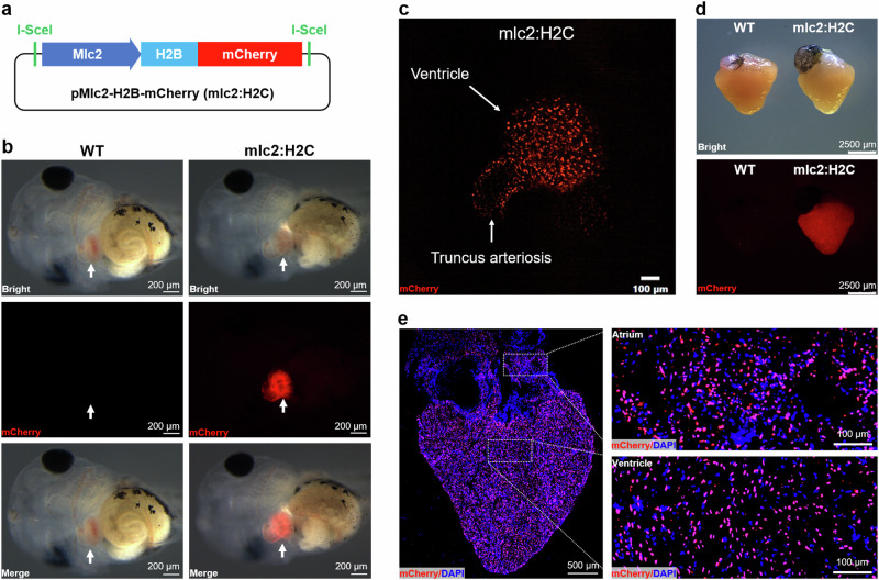
Fig. 1. Generation and characterization of Tg(mlc2:H2C) transgenic X. tropicalis line.
a Schematic representation of the transgenic plasmid pMlc2-H2B-mCherry which harboring the expression cassette of H2B-mCherry (H2C) fusion protein under control of Xenopus mlc2 promoter. b Whole-mount bright-field (upper), epifluorescence (middle), and merged (lower) images showing mCherry expression in Wild-type (WT, left) and F1 Tg(mlc2:H2C) (right) tadpoles. Arrows indicate heart location. c Representative image of the living heart in F1 Tg(mlc2:H2C) tadpole. Red color means mCherry expression specifically in cardiomyocyte nuclei. d Whole-mount bright-field (upper) and epifluorescence (lower) images showing mCherry expression in the adult hearts from WT (left) and F1 Tg(mlc2:H2C) (right) frogs. e Immunostained images for mCherry (red) expression in the adult heart from F1 Tg(mlc2:H2C) frog. DAPI was used as a nuclear stain (blue). Left panel, whole cardiac section in low-magnification. Right panels, high-magnification images of the atrial (upper) and ventricular (lower) regions white boxed in the whole cardiac section. This figure was created by Photoshop Image 12 software using our own data in the work.

