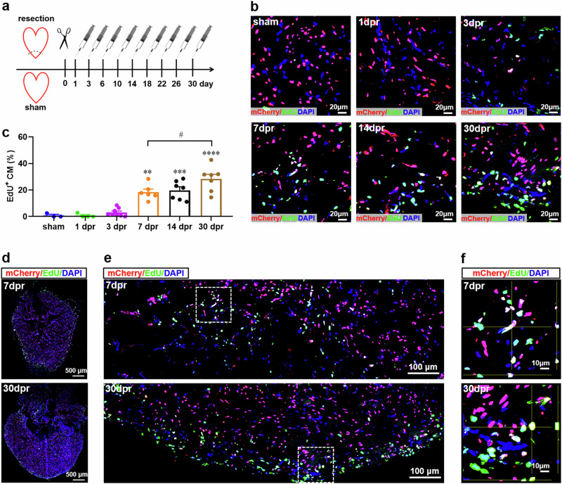
Fig. 5. Cardiomyocyte proliferation contributes to the newly formed myocardium in apex during heart regeneration in adult Tg(mlc2:H2C) transgenic X. tropicalis line.
a Schematic of EdU multiple injection experiment designed to label the proliferating cardiomyocytes in adult Tg(mlc2:H2C) transgenic X. tropicalis line during heart regeneration within 30 days. b Immunostaining for mCherry (red) and EdU (green) expression in the apex of adult hearts from F1 Tg(mlc2:H2C) frogs at the indicated time points after resection. DAPI was used as a nuclear stain (blue). c Quantification of mCherry+EdU+ cells in ventricular apex during heart regeneration within 30 days. Data are presented as mean ± SEM (n = 5 ~ 8 hearts per group). **p < 0.01, ***p < 0.001, ****p < 0.0001 versus sham (one-way ANOVA test). #p < 0.05 (Student’s t test). d Immunostaining for mCherry (red) and EdU (green) expression in a whole cardiac section from the adult heart of F1 Tg(mlc2:H2C) frog at 7 and 30 dpr. DAPI was used as a nuclear stain (blue). e Representative images of mCherry+EdU+ cells in the apical region at 7 and 30 dpr.. f Magnified Z-stack confocal images of mCherry+EdU+ cells white boxed in figure e. This figure was created by Photoshop Image 12 software using our own data in the work.

