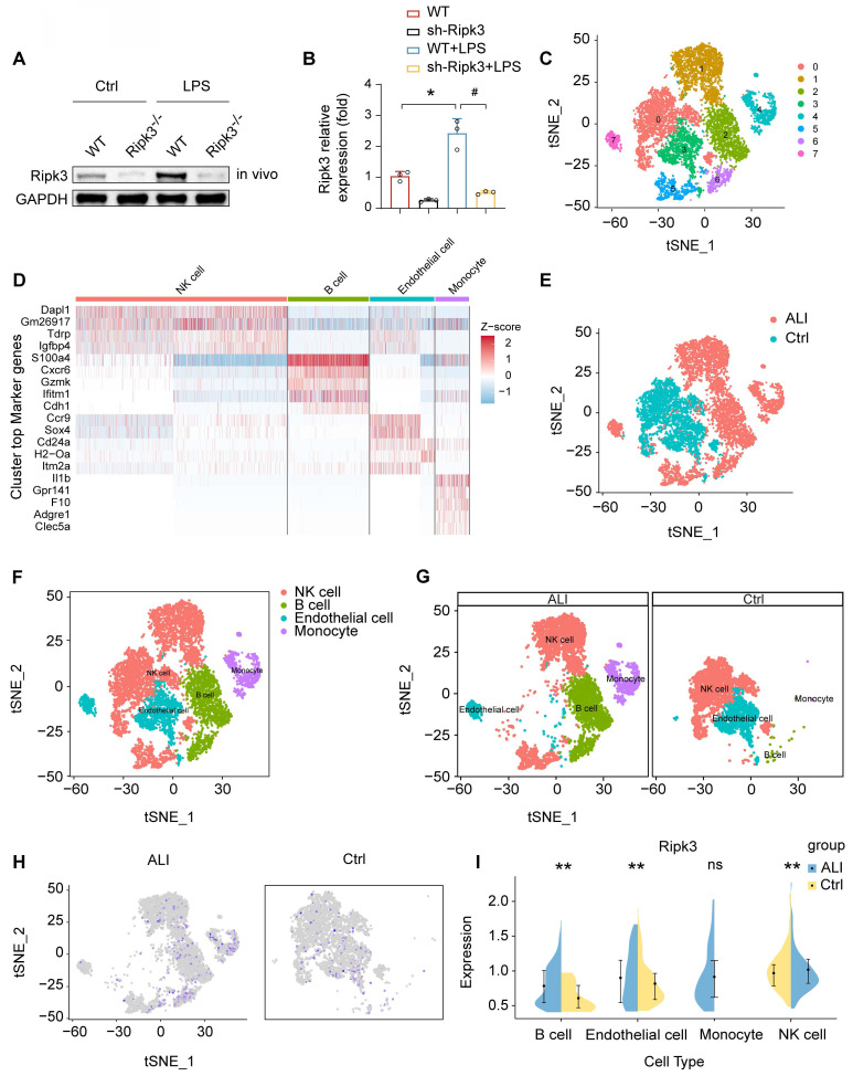
Figure 1.
LPS upregulates Ripk3 in lung ECs. (A-B) The change of Ripk3 expression in vivo was quantified using western blotting. (C-I) Ripk3 expression in different cell types from the lung was determined using single-cell sequencing analysis. Mean ± SEM, ∗p < 0.05 vs. the wild-type (WT) group; #p < 0.05 vs. the LPS group.

