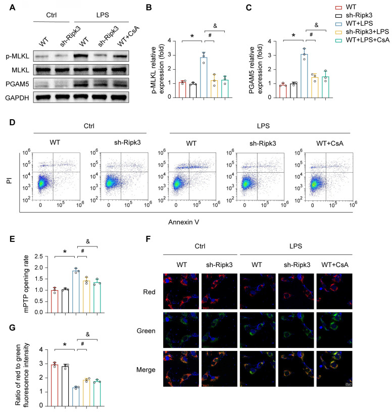
Figure 4.
Ripk3 knockdown reduces PMVEC necroptosis by inhibiting mPTP opening. (A-C) Western blots were used to analyze the expression of PGAM5, and p-MLKL. (D) Necroptosis of PMVECs was quantified by flow cytometry with Annexin V/PI staining. The necroptosis group: the percentage of PI+ cells. Cyclosporin A (CsA) was used as the inhibitor to prevent mitochondrial permeability transition pore (mPTP) opening. (E) Evaluation of the rate of mPTP opening. (F-G) Changes of mitochondrial membrane potential were identified using JC-1 staining. Mean ± SEM, ∗p < 0.05 vs. the Ctrl group; #p < 0.05 vs. the LPS group; &p < 0.05 vs. the LPS+CsA group.

