
Figure 8.
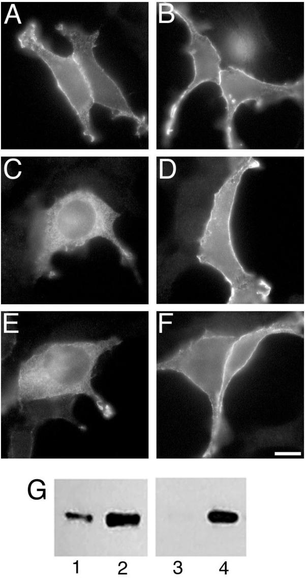
Localization of αi2, sαi2, and αi2A327S and the effect of βγ Expression vectors encoding αi2 (A and B), sαi2 (C and D), or αi2A327 (E and F) were transfected alone (A, C, and E) or together with expression vectors for β1 and γ2 (B, D, and F) into BHK cells. Proteins were visualized by immunofluorescence microscopy using the EE monoclonal antibody followed by an Alexa 488 conjugate anti-mouse antibody. Bar, 10 μm. G, EE-αi2-pcDNA3 (lanes 1 and 2) or EE-sαi2-pcDNA3 (lanes 3 and 4) were transfected into COS-7 cells alone (lanes 1 and 3) or with co-transfection of vectors encoding β1 and γ2 (lanes 2 and 4). Immunoblotting with the EE antibody detected αi2 or sαi2. Note that sαi2 expressed alone (lane 3) is very weakly detected and barely visible in this total cell lysate.