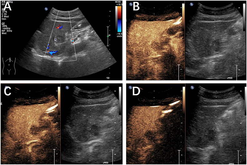
Figure 4.
Histopathology confirmed the presence of a metastatic liver tumor in a 56-year-old female patient with a history of lung cancer. (A) A hypoechoic mass was discovered within the left lobe. On CEUS, the mass exhibited hypo-enhancement during the arterial phase (B), the portal venous phase (60 s) (C), and the late phase (2 min) (D). The lesion was classified as LR-3 based on the CEUS LI-RADS guidelines.

