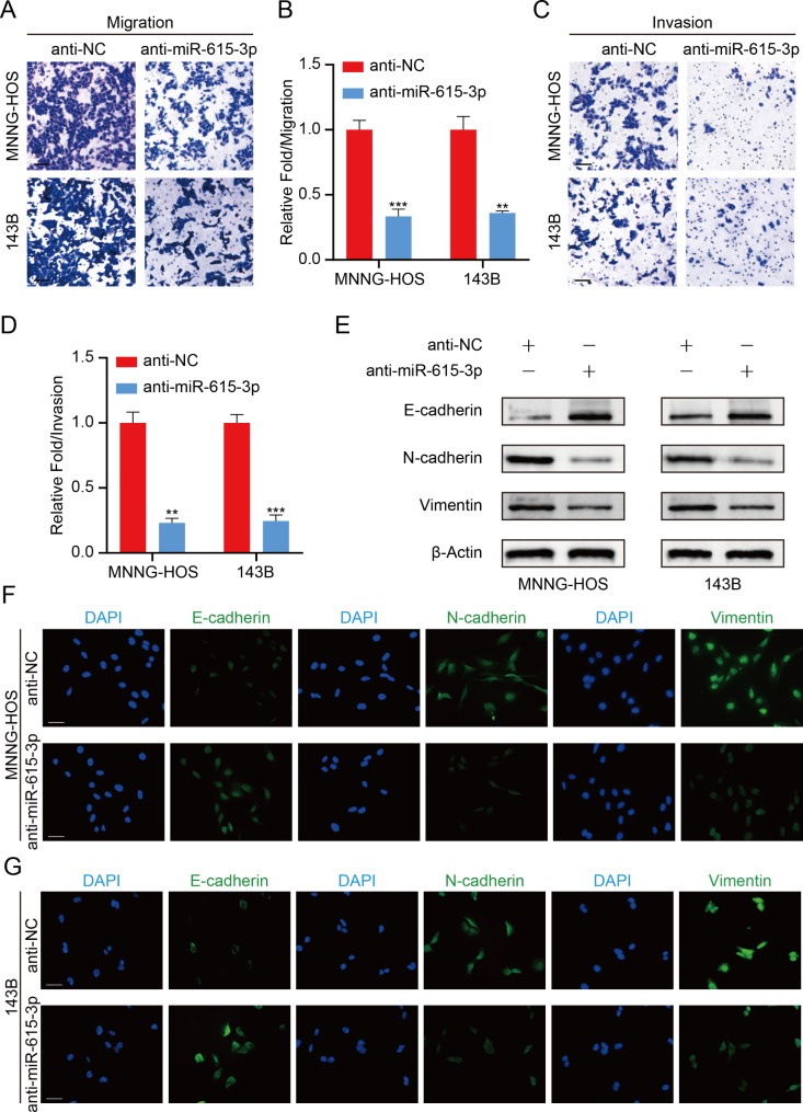
Fig. 3.
miR-615-3p promotes OS cell metastasis via stimulating EMT in vitro. A. Cell migration ability was detected in OS cells with miR-615-3p knockdown or not. Scale bars = 50 μm. B. Quantification of the cell migration ability from the Fig. 3A. C. Cell invasion ability was detected in OS cells with miR-615-3p knockdown or not. Scale bars = 50 μm. D. Quantification of the cell invasion ability from the Fig. 3C. E. Protein levels of EMT markers was determined in different groups (anti-NC and anti-miR-615-3p). F and G. Immunofluorescence staining showed the changes in the expression of EMT markers (green) in OS cells. Nuclei were counterstained with DAPI (blue). Scale bars = 20 μm. Results are displayed as mean ± SD, * indicates p < 0.05, ** indicates p < 0.01, *** indicates p < 0.001

