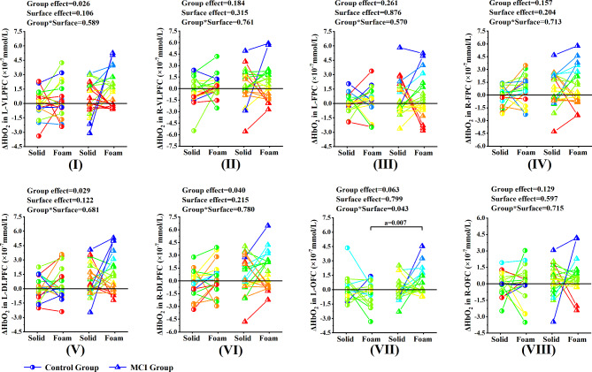
Fig. 5.
Comparison of activation levels of each ROI region between MCI group and control group under the eyes open condition
Note. (I) ~ (VIII) represents the comparison of activation levels of each ROI region between MCI group and control group under the eyes open condition. (I) L-VLPFC, (II) R-VLPFC, (III) L-FPC, (IV) R-FPC, (V) L-DLPFC, (VI) R-DLPFC, (VII) L-OFC, (VIII) R-OFC. L-VLPFC = the left ventrolateral prefrontal cortex and belonged to the left BA45. R-VLPFC = the right ventrolateral prefrontal cortex and belonged to the right BA45. L-FPC = the left frontopolar cortex and belonged to the right BA10. R-FPC = the right frontopolar cortex and belonged to the right BA10. L-DLPFC = the left dorsolateral prefrontal cortex and belonged to the left BA46. L-DLPFC = the right dorsolateral prefrontal cortex and belonged to the right BA46. L-OFC = the left orbitofrontal cortex and belonged to the left BA11. R-OFC = the right orbitofrontal cortex and belonged to the right BA11. a, significant difference between groups. b, significant difference between types of support surface
*p < 0.05, **p < 0.01

