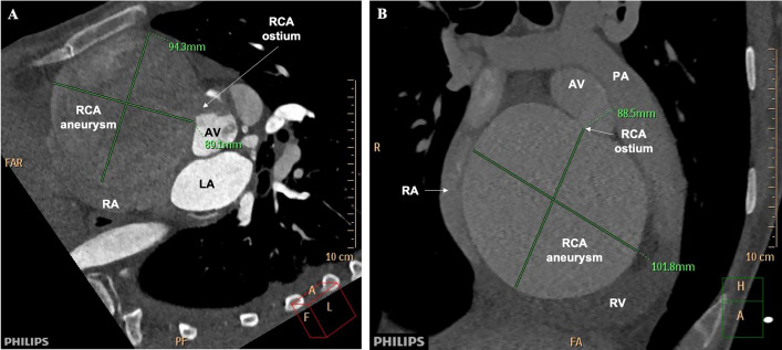
Fig. 3.
Preoperative Computed Tomography Angiography of the aneurysm. A Short axis view of the base of the heart in the arterial phase of contrast injection demonstrating the ostium of the RCA with non-enhanced blood backflow into the aorta and partially enhanced RCA aneurysm compressing the right atrium. B Short axis view at the level of the bifurcation of the pulmonary artery in venous phase demonstrating homogenous enhancement of large RCA aneurysm compressing and distorting the right atrium and ventricle. RV, right ventricle. LA, left atrium. RA, right atrium. TV, tricuspid valve. AV, aortic valve. PA, pulmonary artery. RCA, right coronary artery

