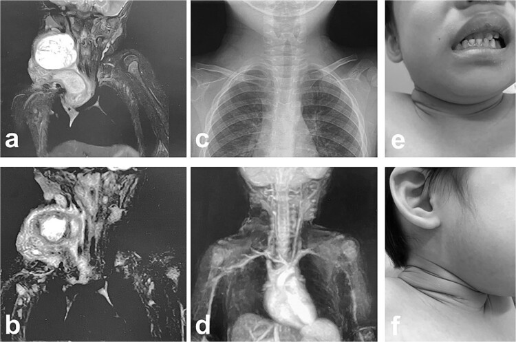
Figure 6.
MRI findings after removal of the Denver shunt catheter. (a) MRI findings showed two different areas of intensity in the neck region and the mediastinal region where the Denver shunt was inserted. (b) MRI findings 2 months postoperatively showed that the mediastinal lesion was smaller with near-complete regression. (c) X-ray showed no tumor shadow and no compression of the trachea by the lymphangioma. (d) MRA showed disappearance of the lymphangioma. (e) and (f) Right cervical swelling did not become evident, even when crying (e: crying, f: smiling).

