
Figure 6. Re-occurrence of amino acids observed in other sarbecoviruses during SARS-CoV-2 evolution.
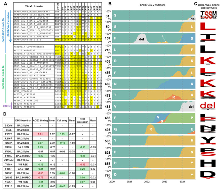
(A) Sequence alignment shows recently mutated amino acids in SARS-CoV-2 that are found in other ACE2-binding sarbecoviruses49. Re-occurring amino acids are highlighted in yellow. Sequence accession codes are provided at the end of each viral strain name. (B) Frequency of amino acids at particular positions on SARS-CoV-2 spike as of October 2024. Data were sourced from nextstrain.org (GISAID data)14,50. (C) Sequence conservation analysis of ACE2-binding sarbecoviruses excluding SARS-CoV-2. Each row corresponds to the residue in panel B. Amino acids that have re-occurred in the currently dominant SARS-CoV-2 variant KP.3.1.1 are shown in red. The SARS-CoV-2 S31 deletion results in an N-glycosylation site 30NFT, corresponding to the N-glycosylation site 30NSS in some other sarbecoviruses, which is also highlighted here. The sequence conservation is plotted by WebLogo51. (D) Fold change in hACE2 binding affinity (Δlog10 KD), entry into 293T cells expressing high ACE2 (functional score), RBD expression (log10(MFI)) for assessment of re-occurring mutations. The average was calculated from all mutations at that site. Green highlights where the effect of the re-occurring mutation is greater than the average, and red highlights where the effect of the re-occurring mutation is less than the average. Data are from previous DMS studies16,41,42. (See also figure S10).