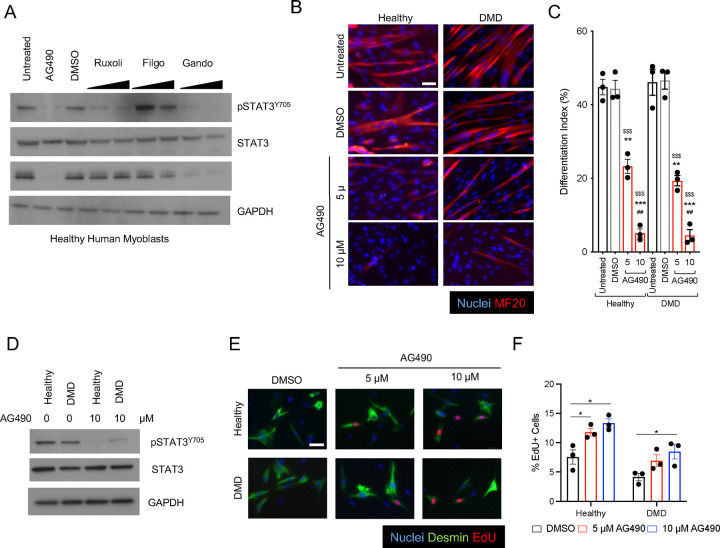
Figure 1: AG490 mediated inhibition of JAK/STAT3 signaling blocks differentiation of human myoblasts.
A. Western blot analysis of whole cell lysate of human healthy myoblasts treated with various JAK inhibitors using a-pSTAT3, a-total STAT3, anti-Myogenin and anti-GAPDH antibodies.
B. Representative immunofluorescence images of myotubes either from healthy or Duchenne patient-derived myoblasts treated with AG490 or DMSO as vehicle control for MF-20 (red), nuclei are counterstained with Hoechst (blue). Scale bar 100 μm.
C. Differentiation index of myotubes for the conditions shown in (B) n=3 biological replicates.
D. Western blot analysis of whole cell lysate of human healthy and Duchenne patient-derived myoblasts treated with 10 μM AG490 using a-pSTAT3, a-total STAT3, and anti-GAPDH antibodies.
E. Representative immunofluorescence images of myoblasts stained with Desmin (green), EdU (red) nuclei are counterstained with Hoechst (blue). Scale bar 20 μm.
F. Quantification of the percentage of proliferating myoblasts treated with DMSO, 5 μM or 10 μM AG490 through EdU staining n=3 biological replicates.
ns. p > 0.05 ; *. p≤ 0.05 ; **. p≤ 0.01 ; ***. p≤ 0.001 unpaired Student’s t test.
* over untreated control; # over lower concentration of drug; $ over DMSO – vehicle control in C.

