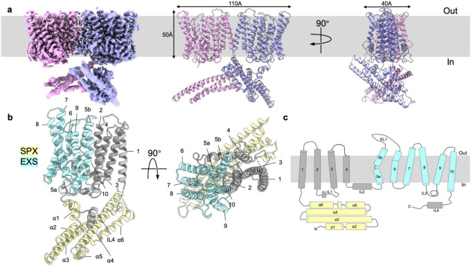
Fig. 1: Overall structure of apo-hXPR1.
a. Cryo-EM density map (left) and cartoon representations of the atomic model (middle and right) of apo-hXPR1 dimer viewed in the membrane plane from two orthogonal directions. Two protomers are colored magenta and lavender. The densities of cytosolic domain and TMD are displayed at a contour level of 8.17σ and 5.04σ respectively. The grey box in the background indicates the membrane bilayer. b. Cartoon representations of an hXPR1 monomer viewed from the side and from top-down. The SPX domain is colored in yellow, EXS domain in light blue, and the rest of the protein in gray. c. Topology of a monomeric hXPR1.

