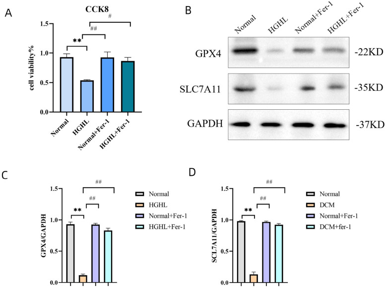
Figure 6.
Ferroptosis involved in HGHL-induced injury in AC16 cells. (A) Viability of AC16 cells evaluated by CCK8 assay. AC16 cells were divided into four groups (Normal, HGHL, Normal + Fer-1, HGHL + Fer-1). Data are expressed as mean ± SD, n = 8 for each group. ** p < 0.01 vs. Normal; # p < 0.05 vs. HGHL; ## p< 0.01 vs. HGHL. (B) Western blots of GPX4 and SLC7A11 in the four groups (Normal, HGHL, Normal + Fer-1, HGHL + Fer-1). (C, D) Quantification of the Western blot results. Data are expressed as mean ± SD, n = 8 for each group. ** p< 0.01, vs. Normal; ## p < 0.01 vs. AC16, human cardiomyocyte cell line. HGHL, high glucose/high lipids. Fer-1, ferrostatin-1.

