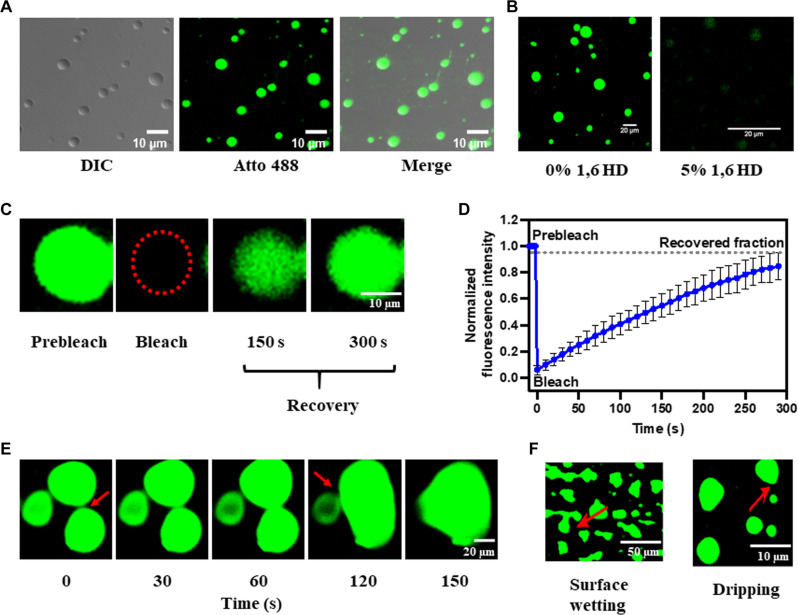
Fig. 2. HuNoV GII.4 RdRp undergoes LLPS in vitro.
(A) Differential interference contrast (DIC) and confocal images of phase-separated RdRp (1% Atto 488 labeled and 99% unlabeled). Phase-separated RdRp has liquid-like properties. (B) Effect of 1,6 HD on LLPS of RdRp. (C) FRAP of RdRp in condensate photobleached at the position indicated by the red circle. (D) The graph shows the recovery curve where normalized fluorescence intensity was plotted against time. (E) Time-lapse images of RdRp condensates exhibit the formation of larger condensates by the fusion of smaller condensates over time. The red arrows indicate the site of fusion. (F) Surface wetting and dripping using confocal microscopy. The red arrows indicate the position of surface wetting and dripping.

