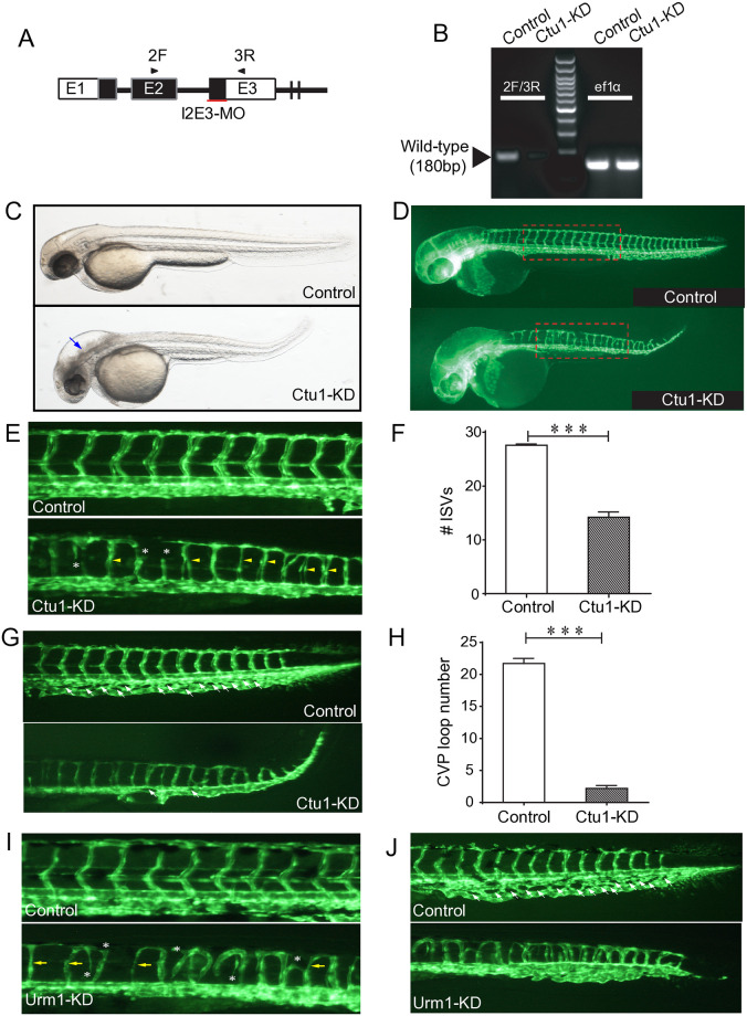
Fig 2. ctu1 morphant zebrafish larvae exhibits developmental defects.
(A) Ctu1-targeted MO design strategy. (B) PCR analysis of control and Ctu1 morphant. (C and D) Bright-fieldand EGFP fluorescentimages depict the overall morphology of control and Ctu1 morphant at 2-dpf. Blue arrows indicate expanded brain ventricle and hindbrain edema in ctu1 morphant compared with control. The dotted square regions are shown at higher magnification in E. (E and G) Image of trunk regions. Compared with control MO, embryos injected with ctu1-i2e3-MO present a lower number of incomplete and thinner intersegmental vessels (ISVs, yellow arrows), and ectopic sprouts (asterisk) of dorsal aorta (E, lower panel). In control embryos, caudal vein plexus (CVP, white arrows) were formed honeycomb-like structures at the tail around 2-dpf (G, upper panel, arrowheads). In contrast, ctu1 deficency resulted in specific defects in CVP formation (G, lower panel, arrowheads). Quantification of the number of complete ISVs (F) and CVP (H). Columns, mean; bars, SEM (n = 10; unpaired student’s t-test; ***, p < 0.001). (I and J) Image of trunk regions. Compared with control MO, embryos injected with urm1-i2e2-MO present a lower number of incomplete and thinner ISVs (yellow arrows), and ectopic sprouts (asterisk) of dorsal aorta (I, lower panel). In control embryos, CVP (white arrows) were formed honeycomb-like structures at the tail around 2-dpf (J, upper panel, arrowheads). In contrast, urm1 deficency resulted in specific defects in CVP formation (J, lower panel, arrowheads).

