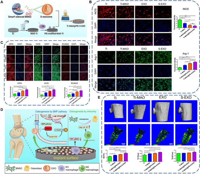
Fig. 3.

(A) Schematic diagram of preparing exosome-functionalized titanium implant by physical adsorption. (B) Immunofluorescence staining images and quantitative results of M1 marker (iNOS) and M2 marker (Arg-1) of macrophages after culturing on the exosome-functionalized titanium surface for 3 d. (C) Immunofluorescence staining images and quantitative results of OPN, OCN, and RUNX2 of BMSCs after culturing on the exosome-functionalized titanium surface for 21 d. (D) Proposed mechanism of the exosome-functionalized titanium implant promoting osteointegration. (E) 3D reconstructed images of micro-computed tomography scanning and quantification of new bone around the implant after implantation for 8 weeks. Reproduced from [125] with permission from the American Chemical Society.
