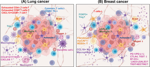
FIGURE 6.

A brief summary of the tumor microenvironment of brain metastasis from lung cancer and breast cancer. Overall, brain metastases from both breast and lung cancers display a is more immunosuppressive and fibrotic microenvironment compared with their primary lesions. (A) Lung cancer. Exhausted T cells, Treg cells, macrophages, neutrophil, microglia, astrocyte, cytokines and chemokines increased. However, dendritic cells, cytotoxic T cells, and B cells decreased. (B) Breast cancer. CCL18+M2 macrophage, RGS5+ fibroblast, myCAF, FOXP3+Treg cells, LGALS1+ microglia, astrocyte increased. And dendritic cells, T cells, and B cells decreased. PD‐1/PD‐L1, CTLA4, TIGIT, B7‐H3 decreased, and the immune checkpoint that mainly mediates immune escape may be related to LAG3–LGALS3 and TIGIT–NECTIN2.
