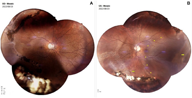
Figure 1.
Preoperative color fundus photographs show (A) an attached retina in the right eye after scleral buckle surgery and (B) an inferotemporal retinal detachment (marked by yellow rhomboids) with a horseshoe tear in the temporal detached retina and an inferonasal attached retina (marked by yellow arrows) in the left eye.

