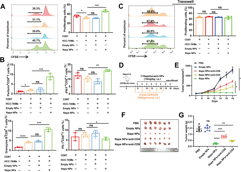
Fig. 8.
Napabucasin-PLGA NPs enhance T cell-mediated anti-HCC immune responses. A–C CFSE-labelled CD8+T cells were activated with anti-CD3 and anti-CD28 antibodies, and then co-cultured with HCC-TAMs pre-treated by Napabucasin-PLGA NPs (3 μM) or Empty NPs in a ratio 4:1 for 72 h. The proliferation of (A) and the production of IFN-γ, Perforin, Granzyme B and PD-1 (B) in these CD8+T cells were measured by flow cytometry. C CFSE-labelled T cells activated with anti-CD3 and anti-CD28 antibodies, and then co-cultured with HCC-TAMs treated as above in Transwell. The proliferation of CD8+T cells was analyzed by flow cytometry. D Experimental scheme for the subcutaneous homograft mouse model in C57BL/6J mice. E The tumor growth curves. F Image of the subcutaneous tumors from each group. G The average tumor weight of each group. n = 6. Napa NPs, Napabucasin-PLGA NPs. Data are shown as mean ± SD, one-way ANOVA with Tukey test. *p < 0.05, **p < 0.01, ***p < 0.001, ****p < 0.0001. ns, no significance

