Fig. 5.
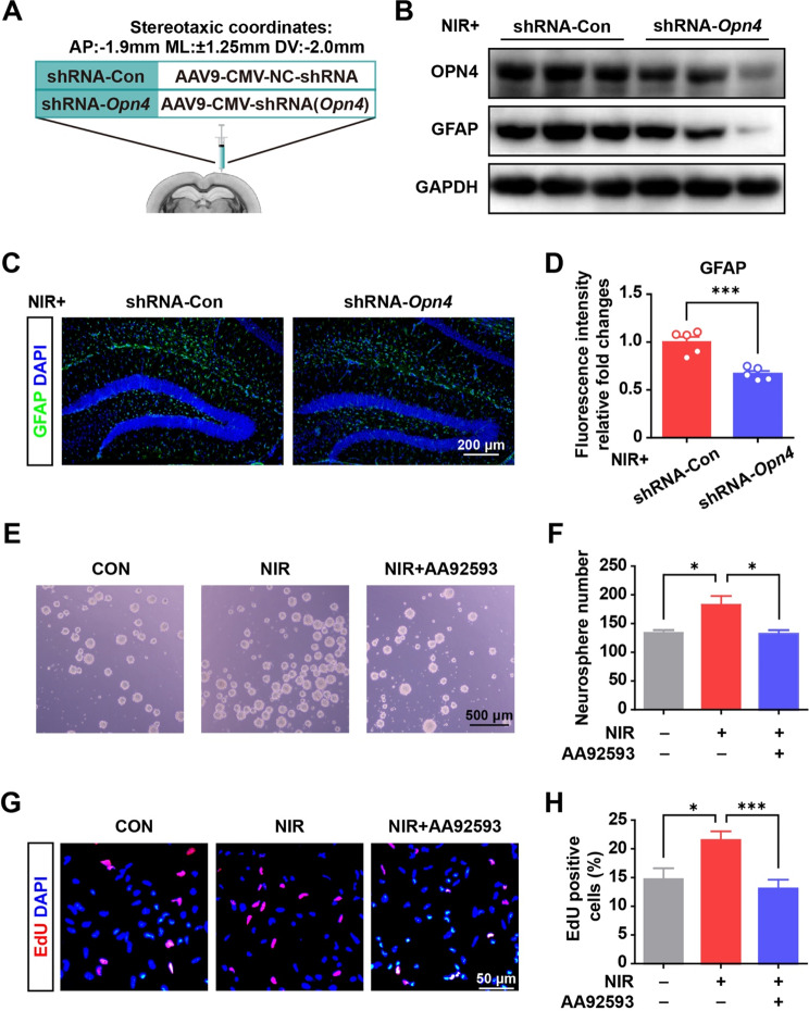
NIR irradiation promoted NSC proliferation and astrocyte differentiation (A) Scheme of the experimental design. (B) shRNA-Opn4 injection down-regulated OPN4 and GFAP expression in the hippocampus of mice with NIR irradiation. (C) Immunofluorescence staining for GFAP in the control and shRNA-Opn4 group with NIR irradiation. (D) Quantitative data from immunofluorescence staining (5 slices from 3 mice for each group). Values are presented as mean ± SEM, ***p < 0.001. (E) Representative micrographs of NSC sphere formation after NIR irradiation and Opn4 inhibitor (AA92593) treatment. (F) Quantitative data from NSC sphere counts with the indicated treatment. The results from four repeat wells of a 24-well plate. Values are given as mean ± SEM. *p < 0.05. (G) Photographs of the EdU incorporation assay of NSCs under NIR exposure. (H) Quantitative data from NSC proliferation under illumination across 12–13 randomly selected view fields of the tested samples using ImageJ software. Values are given as mean ± SEM. *P < 0.05, and ***P < 0.001

