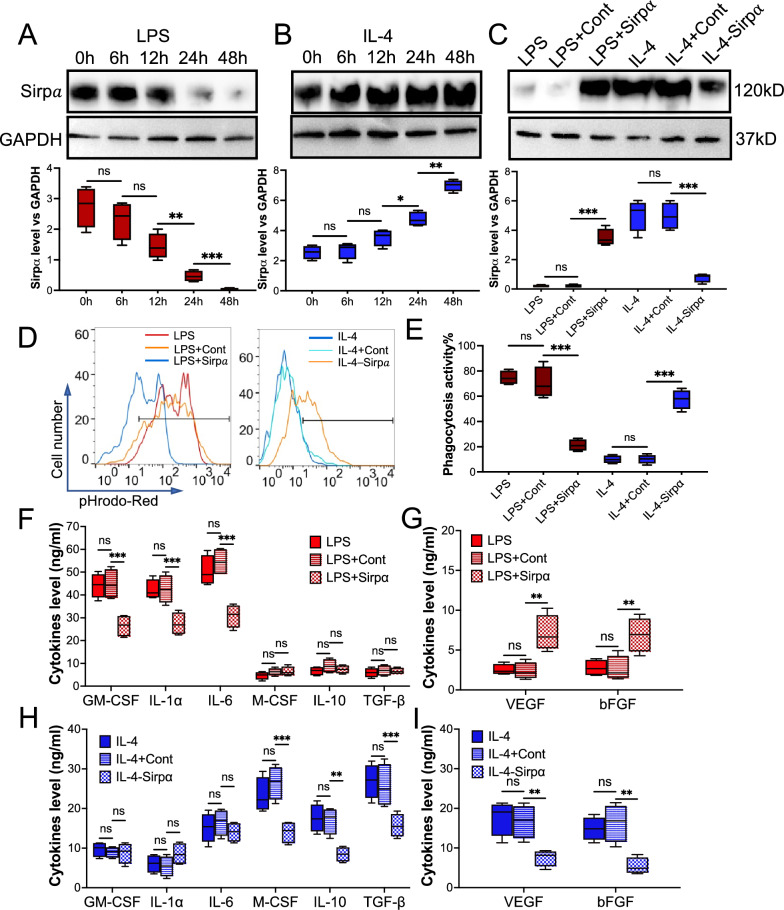
Fig. 3.
Restored or knockdown Sirpα in the macrophage that received LPS or IL-4, respectively, caused function disorder of the macrophage. Macrophages were isolated from the adductor muscles of wild-type mice. Sirpα was increased or decreased in macrophages that pre-treated with 30 ng/ml LPS or IL-4 for 48 h by Lentivirus (LPS + Sirpα: LPS treatment with Sirpα overexpression or IL-4-Sirpα: IL-4 treatment with Sirpα downregulation), respectively. Lentivirus control was used as the control (LPS + Cont: LPS treatment with Lentivirus control and IL-4 + Cont: IL-4 treatment with Lentivirus control). A, B The level of Sirpα (n = 3) in the macrophages that received LPS (A) or IL-4 (B). C The level of Sirpα (n = 3) in the pre-treated macrophages that were incubated with Lentivirus to manipulate Sirpα expression. D, E The phagocytosisof pHrodo Red-labeled apoptotic mCMVECs by the macrophages were measured by flow cytometry (D, total 5000 cells were analyzed). Quantification of the phagocytosis percentages (E). F, G The cytokines (F) and growth factors (G) secreted from the macrophages that received LPS, LPS + Cont or LPS + Sirpα, were measured using ELISA. H, I The secreted cytokines (H) and growth factors (I) from the macrophages that received IL-4, IL-4 + Cont or LI-4-Sirpα. Data is analyzed using one-way ANOVA followed by Tukey multiple range test and expressed as mean ± SD of n = 5, unless specified. *p < 0.05, **p < 0.01 and ***p < 0.001. ns, non-significant

