Fig. 6.
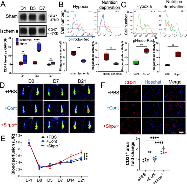
Macrophage from Sirpα knockout (Sirpα−/−) mice promoted angiogenesis in ischemic WT mice at early stage. A The level of CD47 in the adductor muscles of the mouse that received surgery or sham. GAPDH was used as a control. B The phagocytosis of pHrodo Red-labeled apoptotic mCMVECs (hypoxia or nutrition deprivation induced) by the macrophages from the adductor muscles of the mice that received surgery or sham, were tested by flow cytometry. Total 5000 cells were analyzed. C The phagocytosis of pHrodo Red-labeled apoptotic mCMVECs by the macrophages from the adductor muscles of the Sirp−/− mice or WT mice (Cont). D, E The macrophages collected from the adductor muscles of Sirpα−/− mice were transplanted to the ischemic WT mice on day 0 and day 4 post-surgery (early stage), and PBS was used as a control. Laser speckle data showing the relative level of blood perfusion in the hind paws on the indicated days (D, scale bar: 1000 µm). Quantitative analyses of the images showing the left/right ratio of plantar blood perfusion (E). F The mice were euthanized 21 days post-surgery. The sections of the gastrocnemius muscle from the ligated side were subjected to immunohistochemistry analysis for CD31 and counterstained with Hoechst 33342 (E, scale bar: 100 µm). Quantification of the CD31+ area. The CD31+ area on the slide from the mouse administered with PBS was set to 1. Sham: the mice received sham surgery, Ischemia: the mice received femoral artery ligation, + PBS: the ischemic mice received PBS injection, + Cont: the ischemic mice received macrophages from WT mice, + Sirpα−/−: the ischemic mice received macrophages from Sirpα−/− mice. Data is analyzed using one-way ANOVA followed by Tukey multiple range test and expressed as mean ± SD of n = 5, unless specified. *p < 0.05, **p < 0.01, ***p < 0.001 and ****p < 0.0001. ns, non-significant

