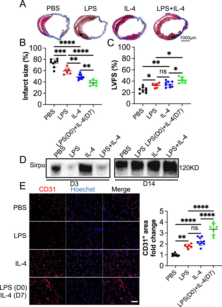
Fig. 8.
LPS and IL-4 worked in a coordinated way to promote angiogenesis and function recovery in the mouse ischemic heart model. Mouse myocardial infarction (MI) was induced by permanently ligating the left anterior descending (LAD) coronary artery. PBS (PBS, n = 8), LPS at early-stage (LPS, n = 6), IL-4 at late-stage (IL-4, n = 9) or LPS at early-stage plus IL-4 at late-stage (LPS (D0) + IL-4 (D7), n = 7) were tail vein injected, respectively. Four weeks post-surgery, cardiac function was evaluated with echocardiography, and hearts were collected thereafter for morphological examination. A and B Infarct size was evaluated with the fibrotic area of the left ventricle by using Masson’s trichrome staining. The blue color represents fibrosis (scale bar: 1000 µm). C Left ventricular fractional shortening (LVFS) was determined with echocardiography. D The level of Sirpα in the infarcted heart was determined by using immunoblotting (n = 4). E The sections of the hearts were subjected to immunohistochemistry analysis of CD31 and counterstained with DAPI (scale bar: 100 µm). The CD31+ area in the PBS group was set to 1. Data is analyzed using one-way ANOVA followed by Tukey multiple range test and expressed as mean ± SD. *p < 0.05, **p < 0.01, ***p < 0.001, and ****p < 0.0001. ns, non-significant

