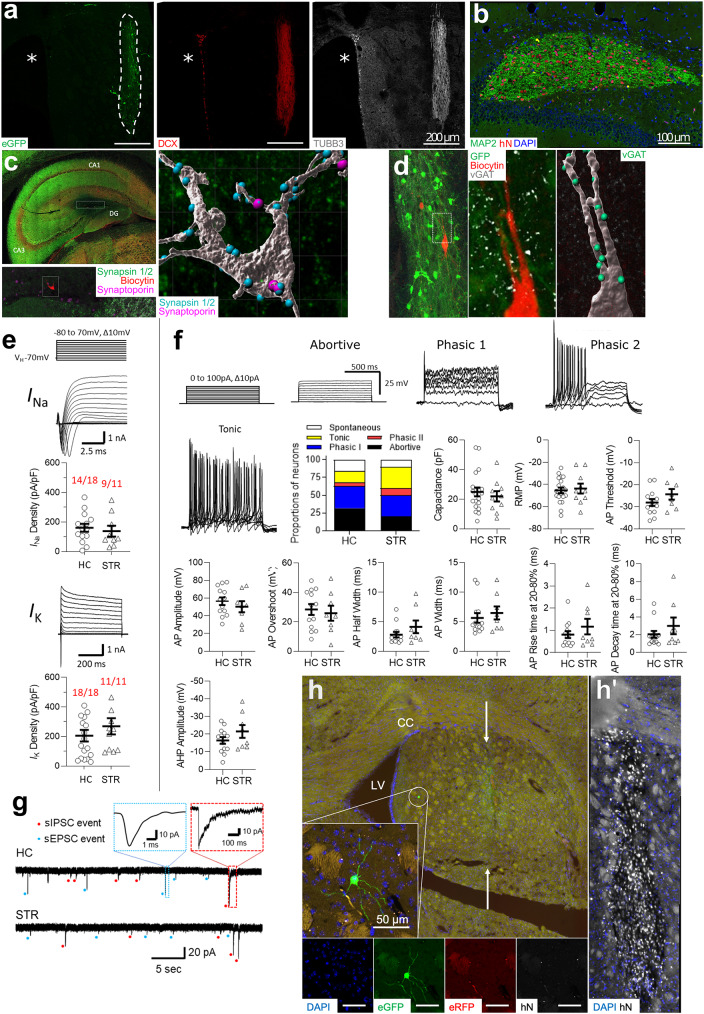
Fig. 1.
Differentiation, functional maturation and integration of engrafted iNSC-derived neurons. (a) STR iNSC graft (dotted line) at 12 weeks after transplantation stained for neuronal markers DCX and TUBB3. Asterisks: lateral ventricle. (b) Ten-week-old iNSC graft in the dentate gyrus labeled with antibodies to human nuclei (hN) and MAP2. (c-d) Staining of presynaptic markers in human neurons, which were identified by their GFP labelling and biocytin content (after biocytin loading during patch clamping). (c) iNSC-derived neuron in a 12-week-old HC graft, which is decorated with synapsin- and synaptoporin-positive puncta, some of which are co-localized. (d) Donor neuron in a 12-week-old STR graft, which is covered with VGAT-positive puncta. The digital reconstruction depicts only puncta in close proximity to the biocytin-labelled neuron (< 1 µm from the surface). (e) Detection of voltage-dependent sodium and potassium currents in engrafted GFP-positive iNSC-derived neurons under voltage clamp condition. Currents were evoked by 500 ms depolarizing voltage steps from − 80 to + 70 mV in 10 mV steps from a holding potential of -70 mV. Potassium currents were measured in the presence of 300 nM tetrodotoxin (TTX). Recordings are from 11 to 24-week-old HC and STR grafts. Red: Number of cells exhibiting sodium and potassium currents vs. number of all measured cells. (f) AP classification in iNSC-derived neurons engrafted into HC (n = 19) and STR (n = 10) at 11–24 weeks after transplantation. (g) Representative spontaneous IPSC and EPSC events that were measured in the presence of TTX in neurons from a 12-week-old HC and a 12-week-old STR graft. These two types of events can be distinguished based on their different release kinetics. (h) RABV-based mono-transsynaptic tracing after infection of a 12-week-old STR graft, depicting a GFP-labeled donor neuron remote from the compact graft core. Note that this cell is negative for the human marker hN and RFP (encoded by donor cells). Insert to the right (h’) depicts the graft core in a consecutive section, labeled with an antibody to hN

