Fig. 5.
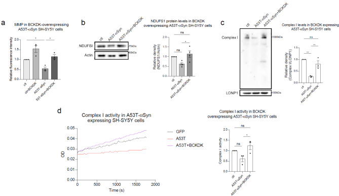
BCKDK expression rescues mitochondrial function in A53T-αSyn cell models. (a) SH-SY5Y cells stably expressing either myc or A53T-αSyn were transfected with BCKDK-myc for 48 h, then stained with TMRM fluorescence dye to measure mitochondrial membrane potential. Fluorescence intensity was quantified per field of view and normalized to the number of cells. Averages for each repeat are shown in the accompanying histogram. n = 3 independent experiments. (b) Total lysates were harvested from stable myc or A53T-αSyn cells transfected with BCKDK-myc for 48 h and subjected to Western blot. Band density of NDUFS1 was normalized to β-Actin (labeled Actin). n = 4 independent experiments. (c) Blue Native PAGE was conducted with mitochondrial fractions to detect Complex I. LonP1, a mitochondrial matrix protein, was used as a mitochondrial loading control. Complex I was marked using an anti-NDUFS6 antibody. n = 3 independent experiments. (d) Complex I activity was assessed using a Complex I enzyme activity assay kit. The resulting slopes were compared as measures of complex activity. n = 3 independent experiments. All data shown are mean ± SEM from at least 3 independent experiments and were analyzed using One-way ANOVA

