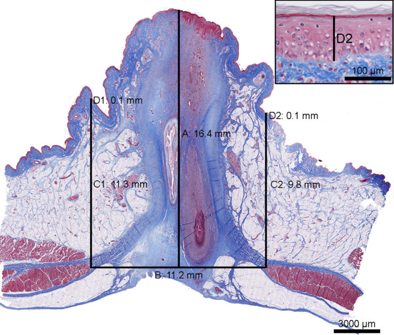
Fig. 1.
Tissue section of the umbilicus from a 14 day old piglet lifted by one hind leg (Group 1). The umbilicus is cut through the centre in the transverse plane, ventrodorsal direction. The thickness of the abdominal wall at the centre (A), the distance between the abdominal muscles (B), the thickness of the abdominal wall at the periphery of the umbilicus (C1 and C2), and the thickness of the epidermis (D1 and D2, insert) were measured. For the measurements C1 and C2 and D1 and D2 the average value was calculated for each. Masson’s Trichrome, scale bar: 3000 μm. Insert: Magnification of D2, scale bar: 100 μm

