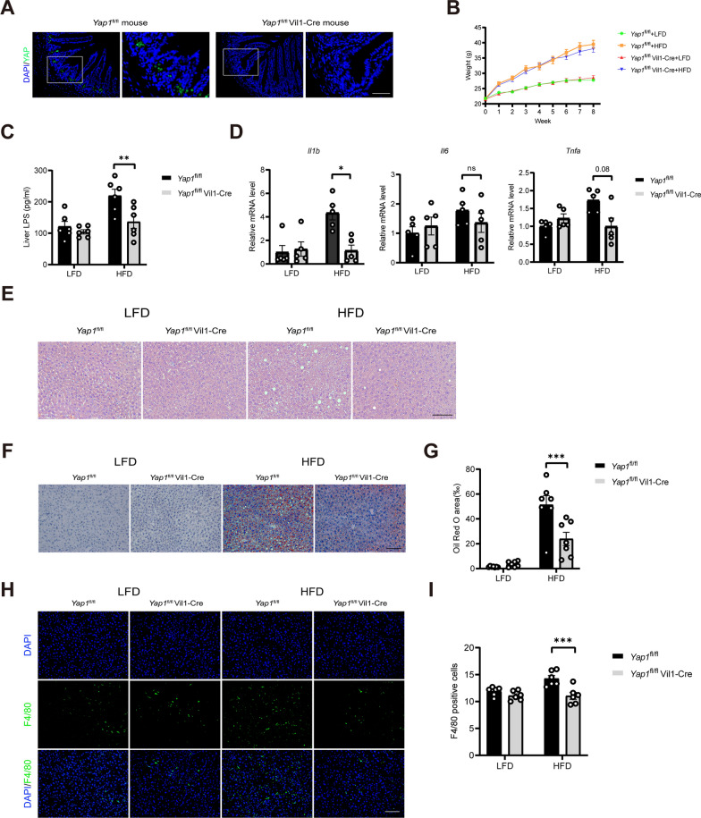
Fig. 2.
An intestinal YAP1 knockout improves hepatic inflammation and steatosis in the HFD-fed mice. A Representative micrographs of immunofluorescence staining for YAP1 in the intestines of the Yap1fl/fl and Yap1fl/fl Vil1-Cre mice. Scale bars: 50 μm. B Body weight changes of the Yap1fl/fl and Yap1fl/fl Vil1-Cre mice in 8 weeks (n = 6). C LPS concentration in the liver tissue of the Yap1fl/fl and Yap1fl/fl Vil1-Cre mice (n = 6). D The mRNA expression of IL-1β, IL-6 and TNF-α in liver tissue of Yap1fl/fl and Yap1fl/fl Vil1-Cre mice (n = 5). E Representative micrographs of H&E staining in liver tissues from the Yap1fl/fl and Yap1fl/fl Vil1-Cre mice. Scale bars: 100 μm. F Representative micrographs of Oil Red O-stained liver tissue from Yap1fl/fl and Yap1fl/fl Vil1-Cre mice. Scale bars: 100 μm. G Quantitative analysis of F (n = 7). H Representative micrographs of immunofluorescence staining for F4/80 in the liver tissues of the Yap1fl/fl and Yap1fl/fl Vil1-Cre mice. Scale bars: 100 μm. I Quantitative analysis of the data in H (n = 6). LFD, low-fat diet; HFD, high-fat diet. *P < 0.05, **P < 0.01, ***P < 0.001, ns: not significant

