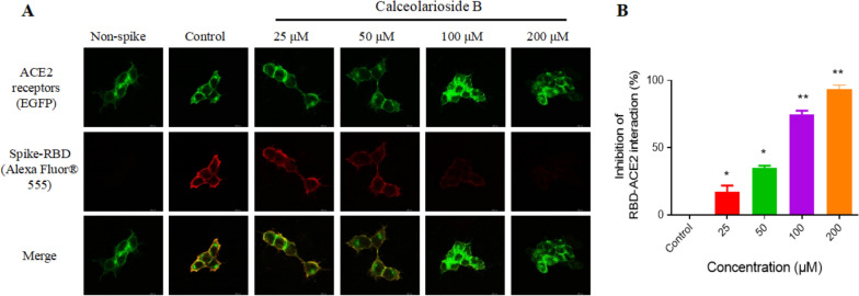
Fig. 3.
Calceolarioside B suppressed the binding of the spike-RBD protein to the ACE2-positive host cells. A Spike-RBD was incubated with calceolarioside B for 1 h and then added to the ACE2-positive cells (green) and incubated for another hour. The protein not bound to ACE2 was washed, and the remaining was labeled with Alexa Fluor® 555 Phalloidin. Fluorescence staining showed reduced binding of spike-RBD (red) to ACE2-positive cells in the presence of calceolarioside B. B The images indicating the spike-RBD–ACE2 binding intensity were quantified by ImageJ. Data were presented as mean ± SD; n = 3; *p < 0.05, **p < 0.01 vs. Control, one-way ANOVA analysis

