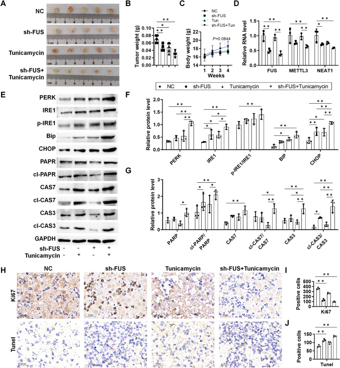
Fig. 6.
In vivo validation of ER stress and apoptosis in GC xenograft mice. sh-NC or sh-FUS-transfected HGC27 cells were inoculated into the armpits of the mice, which were dosed with tunicamycin on days 0, 7, 14, and 21. Tumor tissues were collected on the 30th day. A Tumor tissues harvested on day 30; B tumor volume of the mice; C body weight of the mice measured every week; D relative RNA level of FUS, METTL3, and NEAT1 in the tumor tissues; E western blotting analysis of UPR and apoptosis protein markers; F analysis of UPR markers with ImageJ; G analysis of apoptosis markers with ImageJ; H Ki67, and TUNEL staining of the tumor tissues; I Ki67 positive cells per field; J TUNEL positive cells per field. The cells were counted with ImageJ. N = 3–5 as indicated by each scattered dot. *P < 0.05 and **P < 0.01 by one-way ANOVA followed by Tukey’s multiple comparisons

