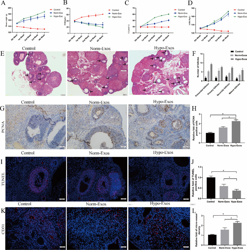
Fig. 4.
Hypoxia pretreatment strengthens the therapeutic angiogenesis effect of hucMSC-exosomes in a rat model of POF in vivo. The POF rats were injected with PBS, norm-Exos and hypo-Exos via the caudal vein. The weights of the rats were measured, and FSH, E2, and AMH serum levels were estimated by ELISA at 0, 1, 2, 3 and 4 weeks after intervention. Histopathological examination of ovarian tissues was performed 4 weeks after treatment. A. The change in body weight was observed in each group. B. The change in FSH serum levels was observed in each group. C. The change in E2 serum levels was observed in each group. D. The change in AMH serum levels was observed in each group. E. HE-stained ovarian sections of POF rats showing the morphologic structure of the ovarian tissues. F. The different developmental stages of follicles were assessed and are shown. G. Representative photomicrographs of PCNA immunohistochemistry staining in ovarian tissues of three groups 28 days after interventions. H. Quantitative analysis of PCNA-positive cells in ovarian tissues. I. Representative photomicrographs of TUNEL staining in ovarian tissues of three groups 28 days after interventions. J. Quantitative analysis of TUNEL-positive cells in ovarian tissues. K. Representative photomicrographs of CD31 immunofluorescence staining in ovarian tissues of three groups 28 days after interventions. L. Quantitative analysis of blood vessel density in ovarian tissues. *P < 0.05 for all figures. n = 10 for each group

