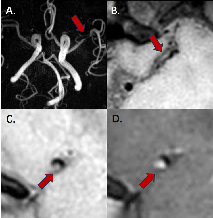
Figure 2.
High-resolution magnetic resonance imaging of a plaque in the left middle cerebral artery.
Notes: (A) Overview of Magnetic Resonance Angiography. The red arrow indicates the narrowest site of the culprit vessel. (B) Overview of the stenosis site at left middle cerebral artery in the pre-contrast T1 axial section. The red arrow indicates the stenosis site. (C) Imaging of the narrowest site at left middle cerebral artery in the pre-contrast T1 sagittal section. The red arrow indicates the culprit plaque at the stenosis site. (D) Imaging of the narrowest site at left middle cerebral artery in the post-contrast T1 sagittal section. The red arrow indicates the culprit plaque at the stenosis site.

