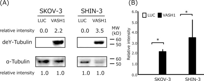
FIGURE 1.

Western blotting of detyrosinated tubulin (deY‐tubulin) in VASH1‐overexpressing or control luciferase gene‐transfected ovarian cancer cell lines. (A) Control cells (SKOV‐3/LUC and SHIN‐3/LUC) very weakly expressed detyrosinated tubulin, while VASH1 gene‐transfected cells (SKOV‐3/VASH1 and SHIN‐3/VASH1) strongly expressed detyrosinated tubulin. α‐tubulin in the cell lysate was used as a loading control. Each number indicates the relative protein expression level compared with α‐tubulin as 1.0. (B) Densitometric analysis is shown in column graph. Data are shown as the mean and SD. *p < 0.05 (vs. LUC gene transfected cells). LUC: luciferase, VASH: vasohibin.
