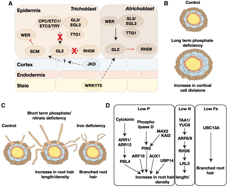
Figure 2.
Genetic pathways controlling root hair plasticity. A) Genes involved in patterning of the root epidermis identified through studies in A. thaliana. In atrichoblast cells, a protein complex consisting of WER, TTG1, and GL3/EGL3 activates the transcription factor GL2 to repress root hair formation. The absence of WER in trichoblast cells results in a protein complex that is unable to activate GL2 to allow root hair development. The patterning of the root epidermis is also influenced by non-cell autonomous functions of JKD (from the cortex) and WRKY75 (from the stele). B)A. thaliana roots exposed to extended periods of phosphate deficient conditions show increased cortical cell divisions. This in turn influences the number of trichoblast cells in the epidermal cell layer, resulting in an increase in root hair number in phosphate-deprived conditions (Janes et al. 2018). C) Adaptation to short-term phosphate and nitrate deficiency results in increased root hair length and density, while iron deficiency causes branched root hairs (Müller and Schmidt 2004). These changes increase the root surface area available for nutrient absorption. D) Auxin and cytokinin signaling pathways influence root hair development under phosphate- and nitrate-deficient conditions. Auxin regulates root hair development through ARF19 in low-phosphate conditions, while changes under low-nitrate conditions depend on ARF6/8. The ubiquitin ligase UBIQUITIN-CONJUGATING ENZYME controls branching of root hairs observed under iron-deficient conditions.

