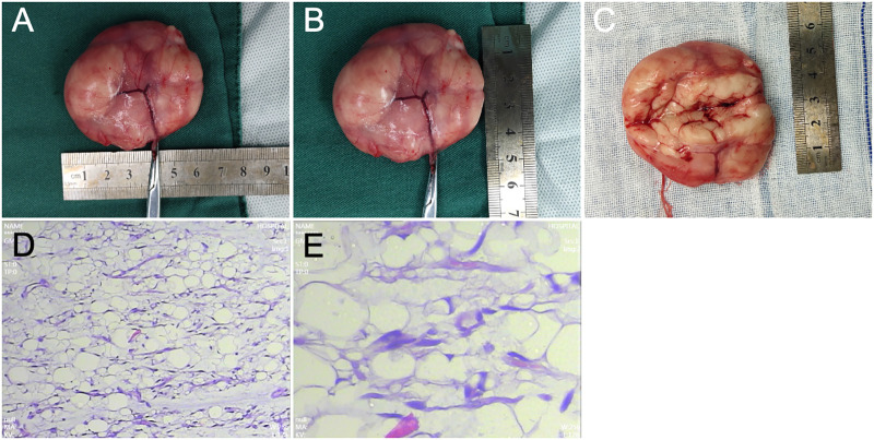
Figure 3.
(A–C) The mass in the right axilla, with a smooth surface and an envelope, measuring 6.0 cm × 4.5 cm × 2.0 cm. The cross section appears gray, solid, and soft, with a pale and uniform texture and an obvious envelope. (D) Under a low-power field-of-view (10×) lens, the tumor is composed of mature fat cells, with slight differences among larger tumor cells; the tumor is divided into irregular lobules by fibrous tissue, with an uneven distribution of capillaries within the tumor tissue. (E) High-magnification field (40×) shows mature white fat cells with flattened eccentric nuclei and thin cytoplasm.

