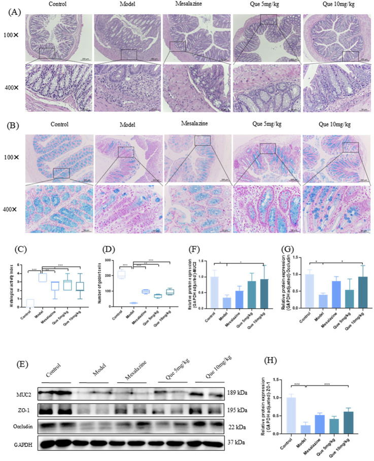
FIGURE 5.
Que restores the integrity of the intestinal mucosal barrier in DSS-induced murine models. (A, C) Hematoxylin and eosin staining of colon tissue sections (×100 and ×400 magnification, respectively) and histopathological scores (n = 4). (B, D) Alcian blue staining (×100 and ×400) of goblet cells in mouse colons. (n = 4). (E–H) MUC2, ZO-1, and Occludin levels in mouse colons (n = 4). *p < 0.05, **p < 0.01, ***p < 0.001.

