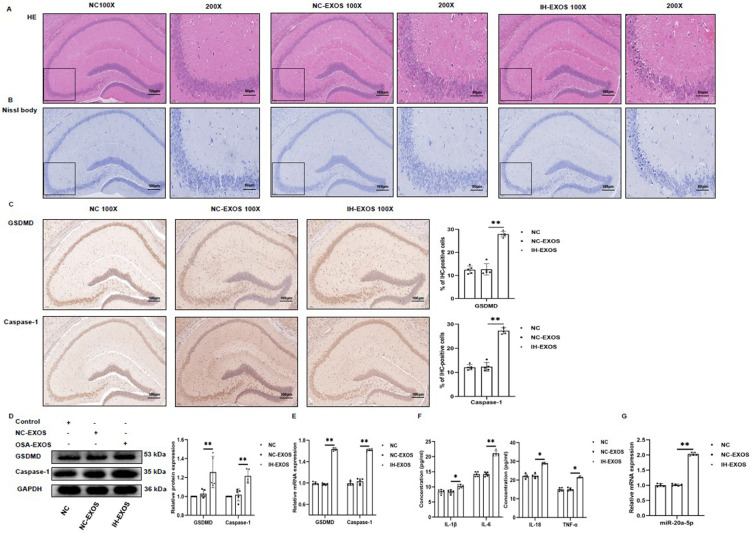
Figure 7.
IH-induced exosomes promote pyroptosis and inflammation in the hippocampus in vivo (n=15). (A) The hippocampus was stained with H&E to assess pathological changes. (B) The hippocampus was stained with Nissl to assess neuronal death. (C) The hippocampus was performed with anti-GSDMD and anti-Caspase-1 for immunohistochemical staining. Magnification×100, the scale is 100 µm. Magnification×200, the scale is 50 µm. (D and E) The expression of GSDMD and Caspase-1 protein and mRNA in the hippocampus was detected by Western blot and qRT-PCR. (F) The levels of IL-1β, IL-6, IL-18, and TNF-α in the serum were detected by ELISA. (G) The expression of miR-20a-5p in the hippocampus was detected by qRT-PCR. Statistical analysis of the data was performed using one-way ANOVA followed by Bonferroni’s multiple comparison tests, df (2, 12). *P<0.05. **P<0.01.
Abbreviation: IHC, immunohistochemistry.

