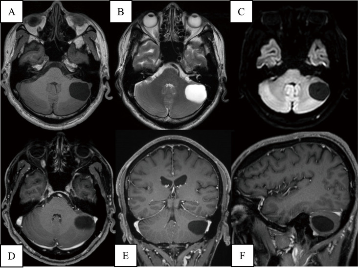
Fig. 1.
Preoperative MRI shows a cystic lesion in the left cerebellar hemisphere. It shows low intensity on T1WI (A), high intensity on T2WI (B), and low intensity on DWI (C). Contrast-enhanced T1WI shows no nodular lesion (D-F). There is no oedematous change in the brain around the mass lesion, and no brainstem compression is found. DWI: diffusion-weighted imaging; MRI: magnetic resonance imaging; T1WI: T1-weighted imaging; T2WI: T2-weighted imaging

