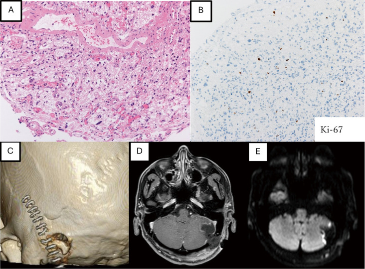
Fig. 4.
Pathological findings show that stromal cells with numerous vacuoles are observed in the stroma, and vascular structures are also observed in the surrounding area (A: Haematoxylin-Eosin, 40×). The Ki-67 positivity rate is 3.1% (B: Immunohistochemistry, 40×). Postoperative imaging, a small craniotomy with a diameter of approximately 2 cm is performed (C). MRI shows that the cyst is reduced in contrast-enhanced T1WI (D), and no high-intensity lesion is observed on DWI (E). DWI: diffusion-weighted imaging; MRI: magnetic resonance imaging; T1WI: T1-weighted imaging

