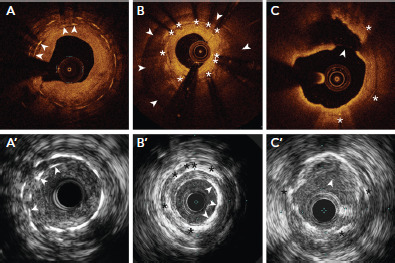
Figure 2: Stent Fracture and Neoatherosclerosis From Different Cases.

In (A) and (A’), the overlapped stent struts (arrowheads) were consistent with stent fracture on optical coherence tomography and intravascular ultrasound (IVUS), respectively. In (B), there is in-stent (white asterisks) restenosis with neointimal hyperplasia. The cause of restenosis is stent underexpansion due to circumferential calcium behind the stent (white arrowheads). In (B’), the stent (black asterisks) is well-expanded with neointimal calcification on IVUS (white arrowheads). In (C), optical coherence tomography shows neointimal rupture in the lipidic plaque within the stent struts (white asterisks). This was not clearly seen on IVUS in (C’). Source: Maehara et al. 2017.[9] Reproduced with permission from Elsevier.
