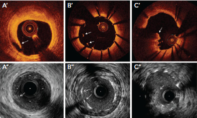
Figure 4: Stent Edge Dissection, Stent Malapposition, and Tissue Protrusion Through Stent Struts on IVUS and OCT.

Medial dissection flap (A’,A’’), stent malapposition (B’,B’’) and tissue protrusion through stent strut (C’,C’’) on optical coherence tomography and intravascular ultrasound, respectively.
Source: Maehara et al. 2017.[9] Reproduced with permission from Elsevier.
