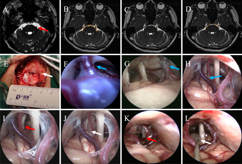
Fig. 1.
Preoperative MRI and surgery of patients with TN underwent fE-MVD. A Picture shows the patient's preoperative 3D FIESTA MRI imaging, with the red arrow pointing to the responsible vessel, which forms a compression in the REZ of TGN; B Area values of bilateral CPA in 3D-FFE sequence axial slices (shown by the yellow line), the area of the healthy (left) side CPA is larger than that of the affected (right) side, and the ratio of CPA area (healthy/affected side) = 1.67/0.91 = 1.84 > 1; C Length values of bilateral TGN in 3D-FFE sequence axial slices (shown by the yellow line), it can be seen that the length of the TGN of the healthy (left) side is longer than that of the affected (right) side, and the ratio of the length of the TGN (healthy/affected side) = 0. 82/0.45 = 1.82 > 1; D TGN pinch angle value in 3D-FFE sequence axial slices (shown by the yellow line), it can be seen that the pinch angle on the healthy (left) side is larger than that on the affected (right) side, and the TGN pinch angle ratio (healthy/affected side) = 51.8/42.4 = 1.22 > 1. E Surgical incision and bone window. Small bone window craniotomy was performed to form an elliptical bone window with a diameter of 2.0 to 3.0 cm, which can clearly expose the junction of transverse sinus and sigmoid sinus (indicated by the white arrow). F–H Neuro-endoscopic classification of PV (indicated by the blue arrow). F Type I: length < 5 mm and the tension is high; G Type II: length between 5 and 10 mm and the tension is moderate; H Type III: length > 10 mm and the tension is low. I–J Pictures show that the responsible artery (indicated by the red arrow) forms a contact compression on TGN from REZ to Meckel's cave, and that the neurovascular decompression is adequate and definitive with the aid of the neuroendoscope with a Teflon cotton placed under direct vision (indicated by the white arrow). K–L Artery and vein are compressed in parallel above the TGN. The red arrow indicates artery and vein, and the white arrow indicates cotton. MRI, magnetic resonance imaging; TN, trigeminal neuralgia; fE-MVD, full-endoscopic microvascular decompression; REZ, root entry/exit zone; TGN, trigeminal nerve; CPA, cerebellopontine angle

