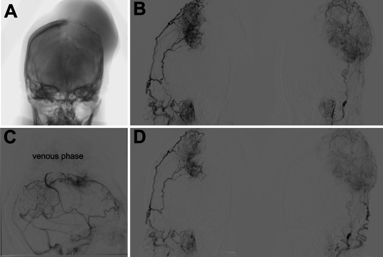
FIG. 2.
Preoperative angiograms. A: Anteroposterior projection showing the location of a large calvarial mass. B: Coronal projection showing extensive bilateral external carotid artery supply. C: Lateral angiogram showing occlusion of the middle third of SSS in the venous phase. D: Significant blood supply remained following bilateral embolization of the middle meningeal arteries.

