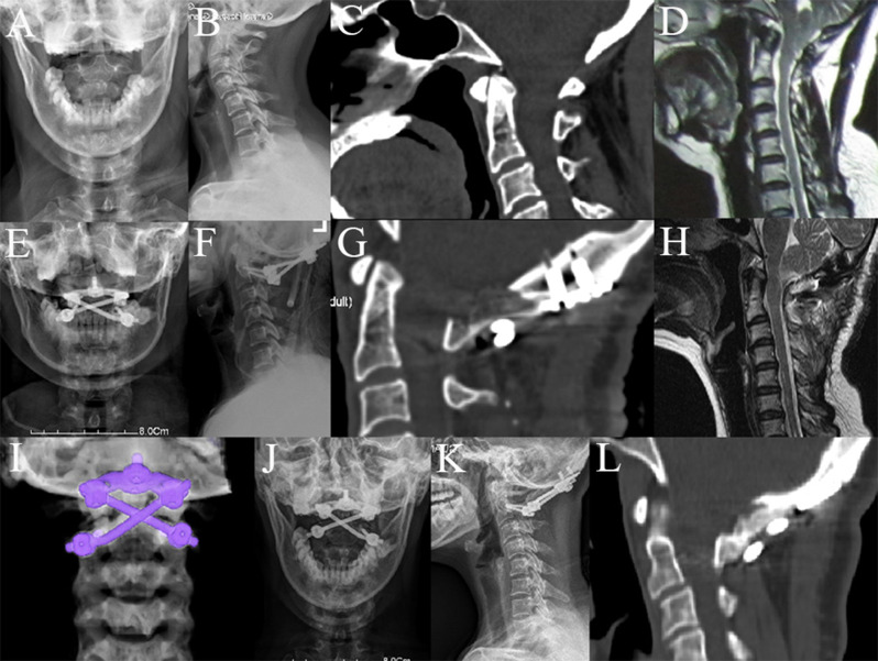
Fig. 2.
A 39-year-old woman diagnosed with basilar invagination and Chiari malformation. A-D. Preoperative X-rays, CT scan and MRI showed evidence of basilar invagination and Chiari malformation. E-I. Postoperative X-rays, CT scan, MRI and three-dimensional reconstruction showed posterior occipitocervical fixation using the crossed rod configuration after C1 laminectomy, enlargement of the foramen magnum, and cerebellar tonsillectomy. J-L. X-rays and CT scan from the 6-month follow-up showed stable fixation and bone fusion

